
Figure 2. Structure of the initial lead and interaction with Hsp90 allosteric site.
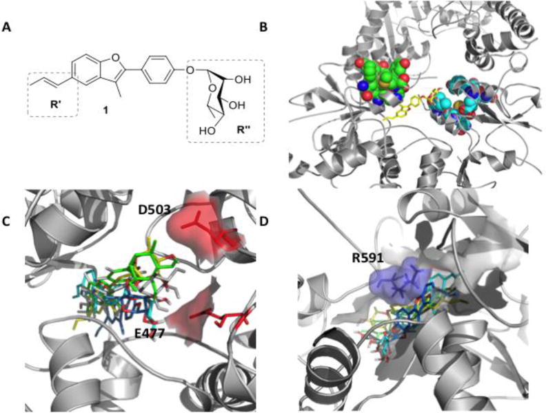
A) Molecular structure of 1. B) 3D structure of compound 1 (yellow) in complex with the closed structure of Hsp90. Van der Waals spheres in light blue and green indicate the client protein Δ131Δ binding site. C) Representative poses of 1 in the representative conformations of the allosteric site, showing the contacts of the ligand with E477 and D503 on protomer A. D) Contacts of 1 with protomer B, highlighting interaction with R591