
Figure 4. Model structures of first-generation allosteric ligands in an ensemble of conformations of the putative allosteric pocket.
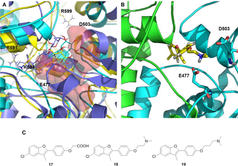
A) Representative structures of the poses of compounds 4 (blue), 10 (cyan) and 12 (yellow). The red surfaces indicate the locations of charged residues targeted by the ligands. B) Representative structure of compound 18 in the allosteric binding pocket. The two Hsp90 protomers are colored differently. C) Compounds 17 to 19.