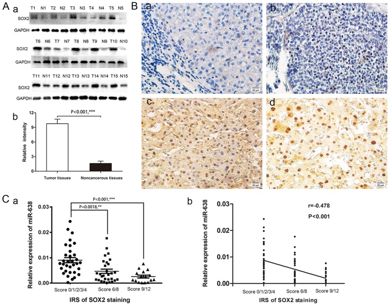
Figure 2.
SOX2 was overexpressed in HCC tissues. (A) Expression of a) SOX2 protein in HCC tissues and matched adjacent non-cancerous tissues in 15 randomly selected patients with HCC was detected by b) western blot analysis. (B) Representative images of immunohistochemical staining of SOX2 in HCC tissues and adjacent non-cancerous tissues are presented in a-d. (Ba) No staining was detected for SOX2 in the blank control group; (Bb) weak nuclear staining of SOX2 in tumor cells; (Bc) medium nuclear staining of SOX2 in tumor cells; and (Bd) strong nuclear staining of SOX2 in tumor cells. (C) The a) semi-quantitative IRS of SOX2 staining was b) negatively correlated with the miR-638 levels in HCC tissues (one-way analysis of variance, P<0.05; Spearman's, r=−0.478; P<0.001). **P<0.05, ***P<0.001.SOX2, sex-determining region Y-box 2; HCC, hepatocellular carcinoma; IRS, immunoreactivity score; miR, microRNA.

