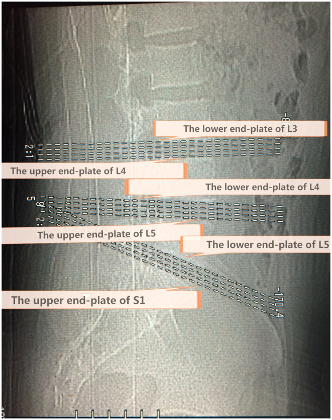
Figure 1.
A locating window view representing the three standardized views positioned accurately along the lower end plate of L3, the upper and lower end plate of L4, the upper and lower end plate of L5, and the upper end plate of S1. Intervertebral segments standardized transaxial images and four levels for each section, positioned accurately through an end plate of a vertebral body. The L4-L5 segment crosses the lower end plate of L4, and the upper end plate of L5, while the L5-S1 segment crosses the lower end plate of L5 and the upper end plate of S1.

