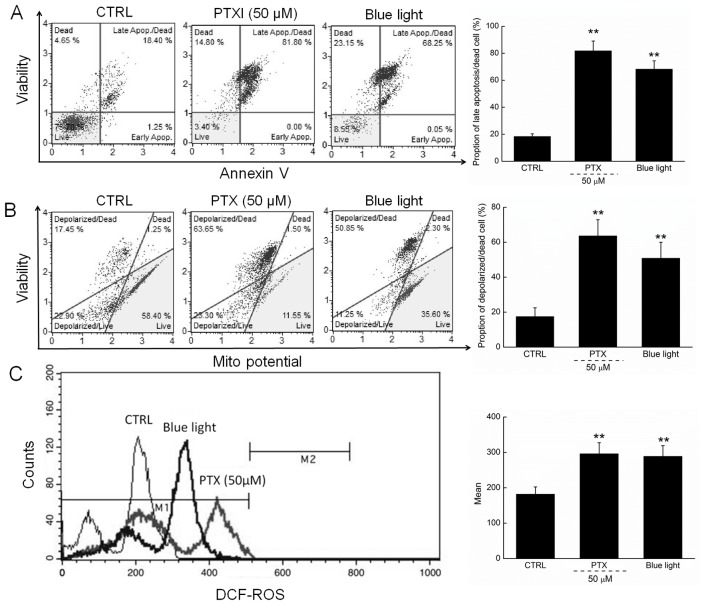
Figure 2.
(A) 24-h exposure to PTX and blue light markedly enhanced the apoptotic rate of HL60 cells. (B) PTX and blue light induced the dissipation of mitochondrial membrane potential, detected via JC-1 staining. (C) 24-h exposure to PTX and blue light significantly enhanced intracellular ROS levels in HL60 cells. Data are expressed as the mean ± standard deviation (n=6). **P<0.01 vs. control cells. PTX, paclitaxel; ROS, reactive oxygen species; JC-1, 5,5′, 6,6′-tetrachloro-1,1′,3,3′tetraethylbenzimidazol-ylcarbocyanine iodide; CTRL, control.

