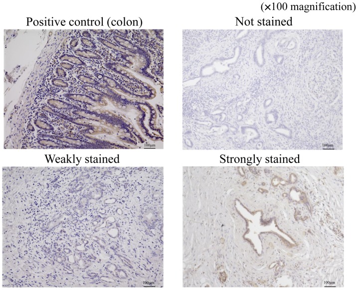
Figure 3.
Immunohistchemistry findings for ferroportin staining are shown at ×100 magnification. Colon sections were used as positive control for ferroportin staining. Stained sections of pancreatic cancer tissues were classified into three categories according to staining intensity: Not stained; weakly stained (stained weaker than positive control); and strongly stained (stained equal to or stronger than positive control).

