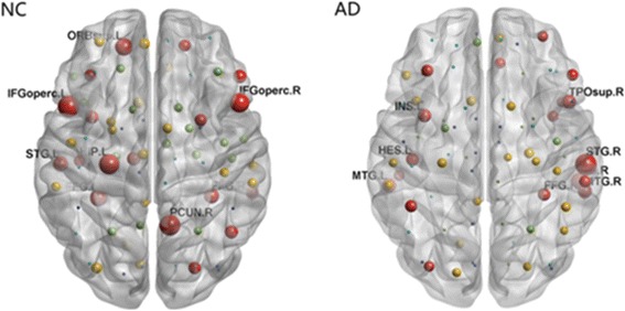
Fig. 3.

Hub regions within groups for MK metric networks. Each ball represented corresponding brain region in AAL atlas, displayed in the center of the region. The size of balls represented the bi value. Only the hub regions with bi>1.5 were indicated by red. Note the hub regions in AD became sparse in the default mode areas. The figure was processed by BrainNet Viewer software. IFGoperc.L: Left inferior frontal gyrus, opercular part; PCUN.R: Right precuneus; IFGoperc.R: Right inferior frontal gyrus, opercular part; STG.L: Left superior temporal gyrus; STG.R: Right superior temporal gyrus; FFG.L: Left fusiform; FFG.R: Right fusiform; ORB.sup.L: Left superior frontal gyrus, medial orbital; HIP.L: Left hippocampus; MTG.L: Left middle temporal gyrus; MTG.R: Right middle temporal gyrus; TPOsup.R: Right temporal pole, superior temporal gyrus; INS.L: Left insula; HES.L: Left heschlgyrus
