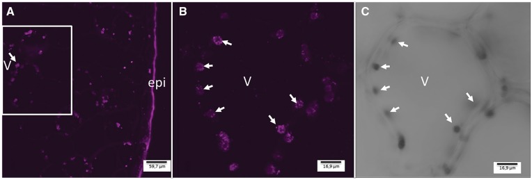
Fig. 6.
Immunohistochemical localization of VpVAN in cytoplasmic organelles in a 7-month-old V. planifolia pod as observed by confocal microscopy. (A) Transverse section of a 7-month-old V. planifolia pod providing an overview of VpVAN localization in the mesocarp. (B) Close-up of a single mesocarp cell showing the intracellular localization of VpVAN in plastids in the cytoplasm. (C) The same section as shown in (B) observed with translucent light. Images were obtained from transverse section of a 7-month-old V. planifolia pod using the VpVAN C-terminal antibody and a goat anti-rabbit antibody labeled with FITC. The images were recorded using a Leica SPII confocal scanning microscope. Arrows indicate the position of selected cytoplasmic plastids harboring VpVAN. Abbreviations: epi, epicarp; v, vacuole.

