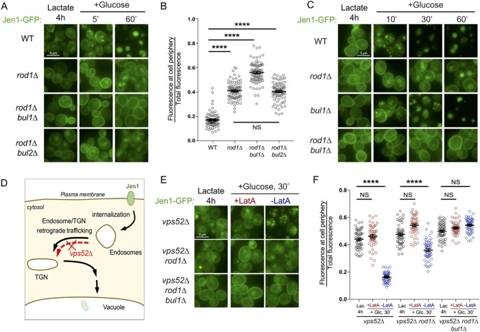
FIGURE 1:
Bul1 assists Rod1 in the glucose-induced endocytosis of Jen1, and acts primarily at the plasma membrane. (A) Localization of Jen1-GFP in WT, rod1∆, rod1∆ bul1∆, and rod1∆ bul2∆ cells after 4 h growth in lactate medium (Jen1 induction) and at various times after the addition of glucose. (B) From the experiment presented in A, quantification of the ratio of fluorescence at the cell periphery over total fluorescence at 60 min after glucose treatment (see Materials and Methods). (C) Localization of Jen1-GFP in WT, rod1∆, bul1∆, and rod1∆ bul1∆ cells after 4 h growth in lactate medium and after glucose treatment. (D) Model of Jen1 trafficking after internalization. The deletion of VPS52 abrogates the vacuolar targeting of Jen1. (E) Localization of Jen1-GFP in vps52∆, vps52∆ rod1∆, and vps52∆ rod1∆ bul1∆ cells grown in the indicated conditions. LatA: latrunculin A. (F) From the experiment presented in E, quantification of the ratio of fluorescence at the cell periphery over total fluorescence at 30 min after glucose treatment.

