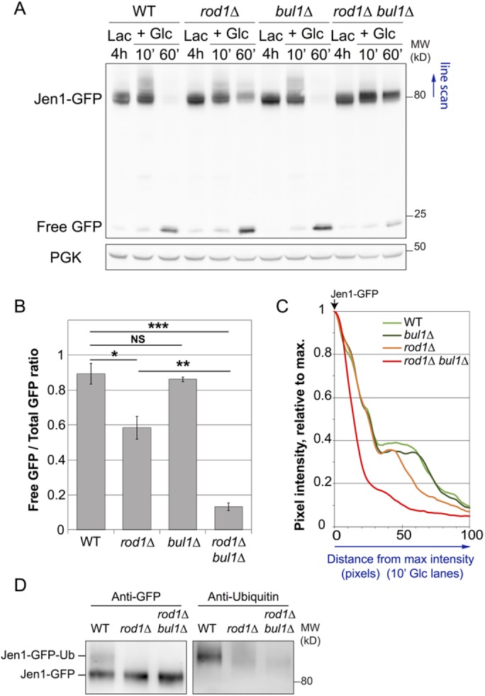
FIGURE 2:

Bul1 is responsible for the residual ubiquitylation and degradation of Jen1 observed in the rod1∆ strain. (A) Degradation of Jen1-GFP over time after glucose treatment in WT, rod1∆, bul1∆, and rod1∆ bul1∆ cells. Total protein extracts were analyzed by SDS–PAGE and Western blotting using the indicated antibodies. PGK: phosphoglycerate kinase (loading control). (B) Quantification of the degradation of Jen1-GFP for each strain (ratio of free GFP over total GFP signal at the 60 min time point; see Materials and Methods). (C) Line scan of the Western blot presented in A showing pixel intensity (relative to the max intensity for each lane) over 100 pixels. (D) Jen1-GFP was immunopurified in denaturing conditions from the indicated strains, 10 min after glucose treatment. Immunoprecipitates were blotted with anti-GFP and anti-ubiquitin antibodies.
