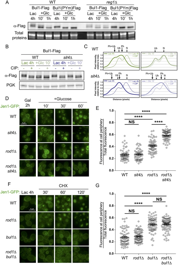
FIGURE 5:
The glucose-induced dephosphorylation of Bul1 requires Sit4. (A) Western blot from total protein extracts prepared from WT or reg1∆ cells expressing either Bul1-Flag or Bul1(PYm)-Flag and grown as indicated. (B) Western blot from total protein extracts prepared from WT or sit4∆ cells expressing Bul1-Flag grown as indicated and treated or not with calf intestinal phosphatase (CIP). (C) Line scan of the Western blot presented in B. (D) Localization of a galactose-inducible Jen1-GFP in WT, sit4∆, rod1∆, and rod1∆ sit4∆ cells after galactose induction or at various times after glucose treatment. (E) From the experiment presented in D, quantification of the ratio of fluorescence at the cell periphery over total fluorescence. (F) Localization of Jen1-GFP in WT, rod1∆, bul1∆, and rod1∆ bul1∆ cells after lactate induction or various times after CHX treatment. (G) From the experiment presented in H, quantification of the ratio of fluorescence at the cell periphery over total fluorescence.

