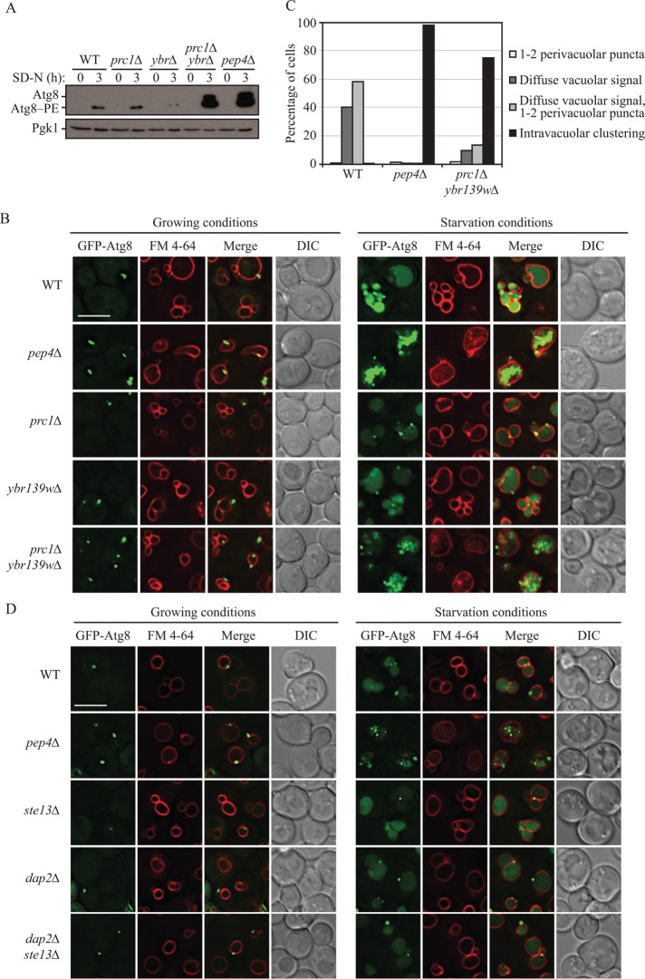
FIGURE 5:
Cells lacking PRC1 and YBR139W are defective in the terminal steps of autophagy. (A) Wild-type (SEY6210), prc1∆ (KPY301), ybr139w∆ (KPY323), prc1∆ ybr139w∆ (KPY325), and pep4∆ (TVY1) cells were grown to mid–log phase in YPD and then shifted to starvation conditions for 3 h. Cells were harvested and protein extracts were analyzed by Western blot using antiserum to Atg8. (B) Wild-type (SEY6210), pep4∆ (TVY1), prc1∆ (KPY301), ybr139w∆ (KPY323), and prc1∆ ybr139w∆ (KPY325) cells expressing GFP-Atg8 from a plasmid were grown in SMD-TRP to mid–log phase. Cells were stained with FM 4-64 for 30 min to label the vacuole and chased in either SMD-TRP for 1 h (growing) or SD-N for 2 h (starvation) before imaging. DIC, differential interference contrast. Scale bar: 5 µm. (C) Quantification of results in B. Cells with GFP-Atg8-positive vacuoles were divided into four categories based on the appearance of the GFP signal as indicated. Wild-type, n = 311 cells; pep4∆, n = 481 cells; prc1∆ ybr139w∆, n = 391 cells. (D) Wild-type (SEY6210), pep4∆ (TVY1), ste13∆ (KPY428), dap2∆ (KPY442), and dap2∆ ste13∆ (KPY443) cells expressing GFP-Atg8 from a plasmid were grown in SMD-TRP to mid–log phase. Cells were stained with FM 4-64 for 30 min to label the vacuole and chased in either SMD-TRP for 1 h (growing) or SD-N for 2 h (starvation) before imaging. DIC, differential interference contrast. Scale bar: 5 µm.

