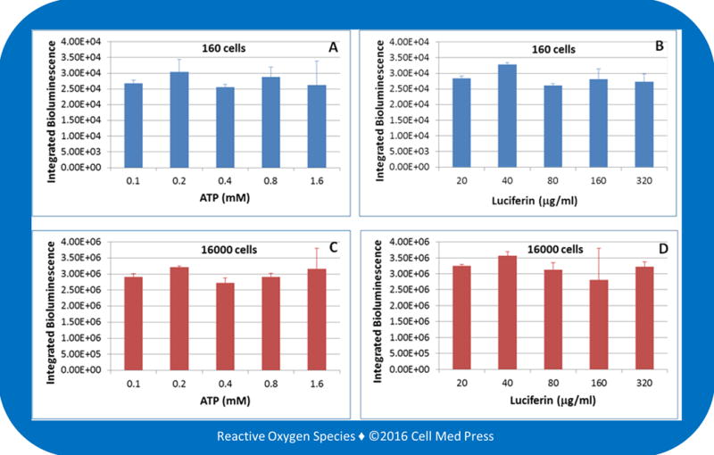
FIGURE 3. Effects of varying the concentrations of ATP and luciferin on B16-F10 melanoma cell-derived bioluminescence.

Bioluminescence was measured at 25.6°C for 5 min after adding samples derived from 160 (panels A and B) or 16,000 (panels C and D) B16-F10-luc-G5 cells (without zeocin selection) to the reaction mix containing 100 μg/ml of luciferin plus the indicated concentrations of ATP (panels A and C) or 0.5 mM ATP plus the indicated concentrations of luciferin (panels B and D). Data represent means ± standard deviation from 4 separate experiments with the unit of bioluminescence being total counts of photon emission over 5 min.
