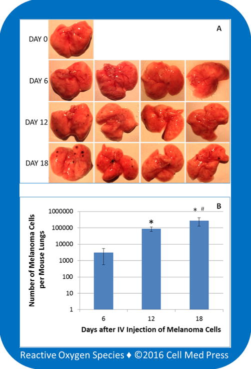
FIGURE 7. Quantification of B16-F10 melanoma cell load in mouse lungs at different time points following intravenous injection of the melanoma cells.

A total of 13 mice were used with one as a control, and each of the remaining 12 mice received 2 × 10⁵ zeocin-selected B16-F10-luc-G5 cells suspended in 0.1 ml of sterile PBS via tail vein injection. Following the intravenous (IV) injection, four mice were euthanized on days 6, 12, and 18, respectively. The lungs were immediately collected for examination of the formation of melanoma foci on the surface of the lungs. After photographing, the entire lungs were homogenized, and 10 μl of the homogenates was used for measurement of bioluminescence in the presence of 100 μg/ml of luciferin and 0.5 mM ATP. The melanoma cell load in each mouse lungs was determined based on a concurrently run standard curve as described in Figure 6. Data represent means ± standard deviation (n = 4).*, p < 0.05 compared with day 6;#, p < 0.05 compared with day 12.
