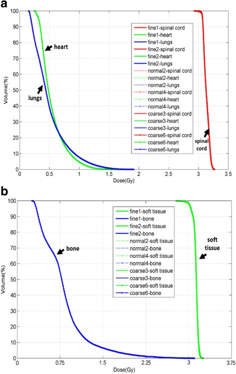
Fig. 3.

DVH for region of interests (ROIs) on chest (Fig. 3a) and pelvic (Fig. 3b) phantoms with the dose calculated on 6 series of MVCT images with arrows pointing to the anthropomorphic ROI in each phantom. The curve names in both legends of these two figures have the same structures. Take ‘fine 1 – spinal cord’ as example: ‘fine’ stands for the acquisition pith (fine, normal and coarse), ‘1’ stands for the reconstruction interval (1, 2, 3, 4 and 6 mm) and ‘spinal cord’ stands for one of the ROIs (spinal cord, heart, lungs on chest phantom, and soft tissue, bone on pelvic phantom) delineated on the MVCT image serials
