Fig. 1.

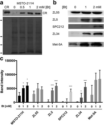
Upregulation of CR in Bt-exposed immortalized mesothelial (Met-5A) and MM cells a Upper panel: CR Western blot of cytosolic proteins from MSTO-211H cells treated with Bt for 72 h; left lane (CR): human recombinant CR (40 ng) as control. Lower panel: Ponceau Red-stained membrane to check for even loading. Ticks on the left mark the position of marker proteins (in kDa, from top to bottom: 75, 35, 28, 10). The signal for CR is slightly above the 28-kDa marker protein. b Western blots for CR of Bt-treated (72 h) cells: one mesothelial cell line (Met-5A) and 4 MM cell lines: ZL55, ZL5, SPC212, ZL34. Equal loading was confirmed by Ponceau Red staining as shown in a (not shown). c Semi-quantitative CR Western blot results (Mean ± S.D.) from 5 independent representative experiments. *** p < 0.001
