Fig. 5.

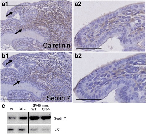
Co-expression of calretinin and septin 7 Immunohistochemical analysis of E10.5 lung tissue stained for CR a1 and a2 and septin 7 b1 and b2. Scale bars: 250 μm for a1 and b1, and 50 μm for a2 and b2. c Left: Septin 7 Western blot of extracts from cultured primary mesothelial cells from C57Bl/6J (WT) and CR−/− mice; note the lower levels of septin 7 in WT cells. Right: Septin 7 Western blot from SV40-immortalzed mesothelial cells derived from WT and CR−/− mice. As loading control (l.c.) an endogenous biotinylated protein (Mr ≈ 75 kDa) was used. The complete Ponceau Red-stained membrane is shown in Additional file 1: Figure S3
