
Figure 6.
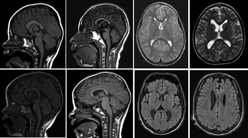
Sagittal T1 MRI scans (left) and axial T2 (top right) and FLAIR (bottom right) MRI Scans depicting common anomalies seen in PTHS including dysplasia of the corpus callosum, bulging caudate, global atrophy, and scattered T2 hyperintensities. Images are from our cohort at UTSW Medical Center / Children’s Health Center for Autism and Developmental Disabilities.