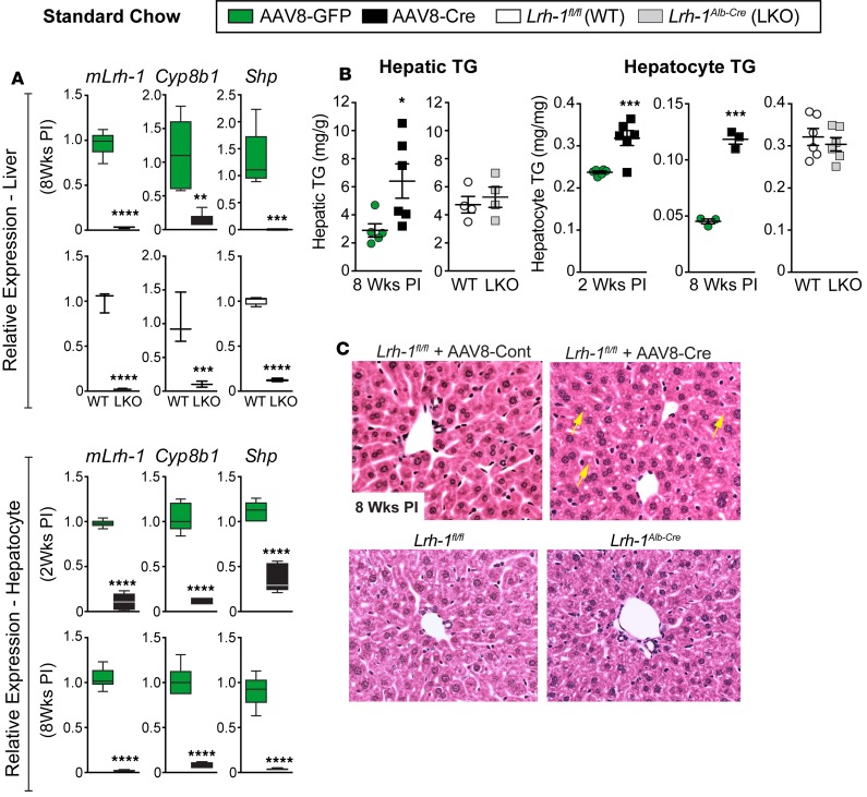
Figure 1. Deleting LRH-1 in adult liver promotes hepatic lipid accumulation.
(A) Relative expression by qPCR analysis of Lrh-1, Cyp8b1, and Shp in livers or primary hepatocytes isolated from Lrh-1AAV8-GFP and Lrh-1AAV8-Cre male mice or from age-matched Lrh-1fl/fl or Lrh-1Alb-Cre mice (n = 5 and n = 6 for Lrh-1AAV8-GFP and Lrh-1AAV8-Cre mice, respectively, and n = 3 for both Lrh-1fl/fl and Lrh-1Alb-cre mice). (B) TG content in livers or primary hepatocytes at two time points after infection (PI) from male mice with groups indicated in the figure. (C) Representative images (original magnification, ×20) of H&E-stained livers from Lrh-1AAV8-Cre and Lrh-1Alb-Cre mice; yellow arrows highlight lipid droplets in Lrh-1AAV8-Cre liver. For primary hepatocytes n = 2 for all genotypes or groups done in triplicate. Error bars represent ± SEM. For box-and-whisker plots, maximum and minimum values are shown with median. *P < 0.05, **P < 0.01, ***P < 0.001, ****P < 0.0001, unpaired Student’s t tests.

