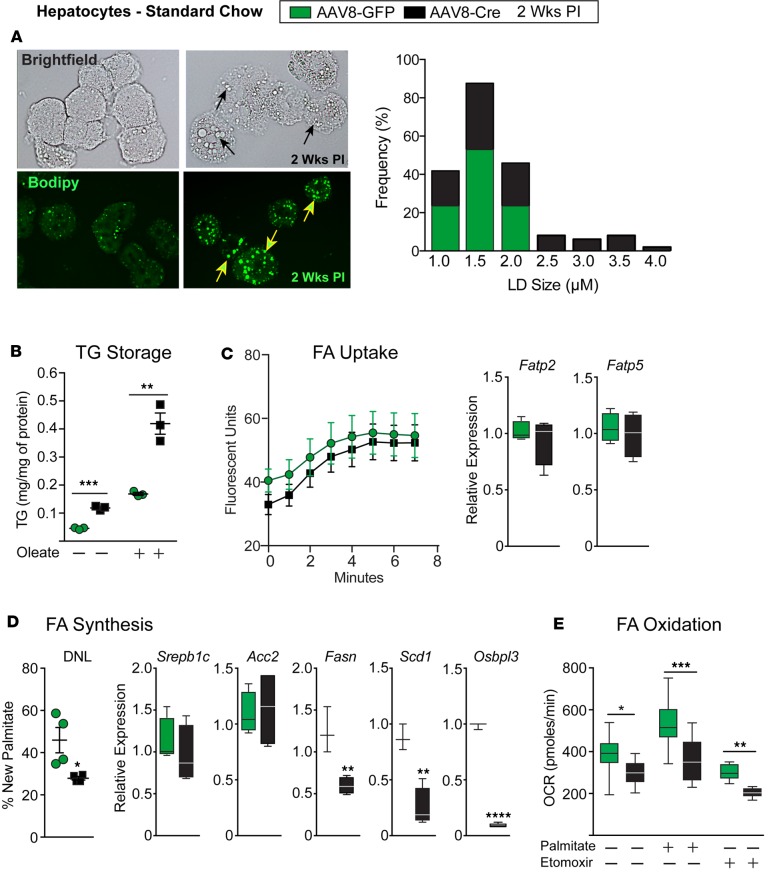
Figure 3. Lrh-1AAV8-Cre hepatocytes preferentially store fatty acids in large lipid droplets.
(A) Representative bright-field images and images of BODIPY-stained primary hepatocytes from Lrh-1AAV8-GFP and Lrh-1AAV8-Cre male mice 2 weeks after infection; black or yellow arrows highlight lipid droplets in Lrh-1AAV8-Cre hepatocytes. Quantification of lipid droplet size and frequency is shown, as measured by ImageJ. (B) TG content obtained from Lrh-1AAV8-GFP and Lrh-1AAV8-Cre mouse hepatocytes with or without FA loading with oleic acid for 16 hours. (C) Measurement of FA uptake and expression of hepatic fatty acid transporters (Fatp2 and Fatp5) in Lrh-1AAV8-GFP and Lrh-1AAV8-Cre hepatocytes. (D) De novo lipogenesis (DNL) in hepatic tissue 2 weeks after infection, as quantified by the percentage of new hepatic palmitate (n = 4 per group), and expression of key lipogenic genes in hepatocytes isolated from Lrh-1AAV8-GFP and Lrh-1AAV8-Cre mice. (E) Fatty acid oxidation in hepatocytes from Lrh-1AAV8-GFP and Lrh-1AAV8-Cre mice. Primary hepatocytes were analyzed from at least 2–4 males per group, with each assay done in triplicate for B–E. Error bars represent ± SEM. For box-and-whisker plots, maximum and minimum values are shown with median. *P < 0.05, **P < 0.01, ***P < 0.001, unpaired Student’s t test for B, D, and E.

