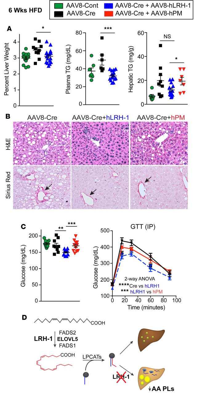
Figure 7. Replacing mouse with human LRH-1 resolves liver injury and improves glucose homeostasis.
(A) Percentage of liver weight and hepatic/plasma TGs in mice fed high-fat diet (HFD) (n ≥ 8 for Lrh-1AAV8-GFP, n ≥ 6 for Lrh-1AAV8-Cre, n ≥ 13 for Lrh-1AAV8-Cre+hLrh1 and Lrh-1AAV8-Cre+hPM, n ≥ 7). (B) Representative images (original magnification, ×20) of H&E- and Sirius red–stained livers from mice fed HFD from different experimental groups. Arrows highlight periportal collagen staining. (C) Fasting plasma glucose in male mice fed HFD for 6 weeks (n = 11 for Lrh-1AAV8-GFP, n = 9 for Lrh-1AAV8-Cre, n = 13 for Lrh-1AAV8-Cre+hLrh1, n = 11 for Lrh-1AAV8-Cre+hPM). Glucose tolerance test (GTT) results (n = 12 for Lrh-1AAV8-Cre, n = 6 for Lrh-1AAV8-Cre+hLrh1, and n = 7 for Lrh-1AAV8-Cre+hPM). (D) Schematic showing that loss of LRH-1 decreases arachidonic acid phospholipids (AA PLs) and leads to hepatic lipid accumulation via regulation of arachidonic acid biosynthesis. Error bars represent ± SEM. *P < 0.05, **P < 0.01, ***P < 0.001, ****P < 0.0001, unpaired Student’s t test (A and C) or 2-way ANOVA with Sidak (C).

