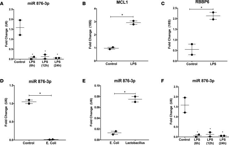
Figure 6. Proteobacteria exposure decreases miR 876-3p expression in vitro.
Normal human bronchial epithelial (NHBE) cells were utilized for in vitro experiments. (A–C) Expression of exosomal miR 876-3p was decreased in supernatants of NHBE cells exposed to LPS at 6, 12, and 24 hours, compared with control (all P < 0.05). A corresponding increase in the target mRNAs MCL1 (P = 0.006 by t test) and RBBP6 (P = 0.03 by t test) were seen at 12 hours. (D) Decrease in exosomal miR 876-3p expression is seen at 12 hours in cell supernatants of NHBE cells exposed to E. coli bacteria (P = 0.002 by t test). (E) Compared with Lactobacillus-exposed cells, E. coli–exposed cells showed decreased exosomal miR 876-3p levels at 12 hours (P = 0.0087 by t test). (F) NHBE cells exposed to normoxia or hyperoxia, with either LPS or saline (control) for 12 hours. Exosomal miR 876-3p expression was decreased in hyperoxic cell supernatants. A greater magnitude of decrease was seen with the addition of LPS in both normoxia and hyperoxia (saline normoxia vs. LPS normoxia, P < 0.0001 by ANOVA; saline hyperoxia vs. LPS hyperoxia, P = 0.03 by ANOVA). *P < 0.05.

