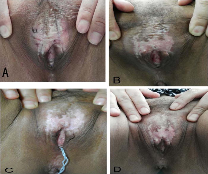
Fig. 1.

The macroscopy characteristics of vulvar lesions of one patient was received three times of ALA-PDT and followed-up in 6 months. (A) Pre-treatment. (B) One-month follow-up. (C) Three-month follow-up. (D) Six-month follow-up. These pictures show a significant clinical improvement with reduction of hyperkeratosis and decrease of the area of vulval lesions, even hypopigmentation was improved.
