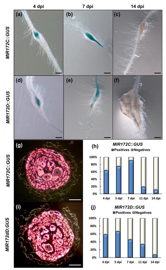
Fig. 4.

A timeline of miRNA172c and miRNA172d promoters indicated early activation patterns after root-knot nematode infection. (a–f) Representative pictures of GUS assays at 4, 7 and 14 d post infection (dpi), respectively of (a–c) MIR172C ::GUS and (d–f) MIR172D -::GUS lines in Arabidopsis galls induced by Meloidogyne javanica. (g, i) Dark field images of Araldite® cross sections of GUS stained galls at 7 dpi showing signal in the giant cells and adjacent cell layers within the vascular cylinder. Asterisks, giant cells; N, nematode. (h, j) Percentage of blue galls for (h) MIR172C ::GUS and (j) MIR172D ::GUS. Bars: (a–f) 200 μm; (g–i) 50 μm.
