
Figure 1. MsrB3 protein signal in hippocampal pyramidal layer, vascular walls, choroid epithelium, and ependymal ventricular lining.
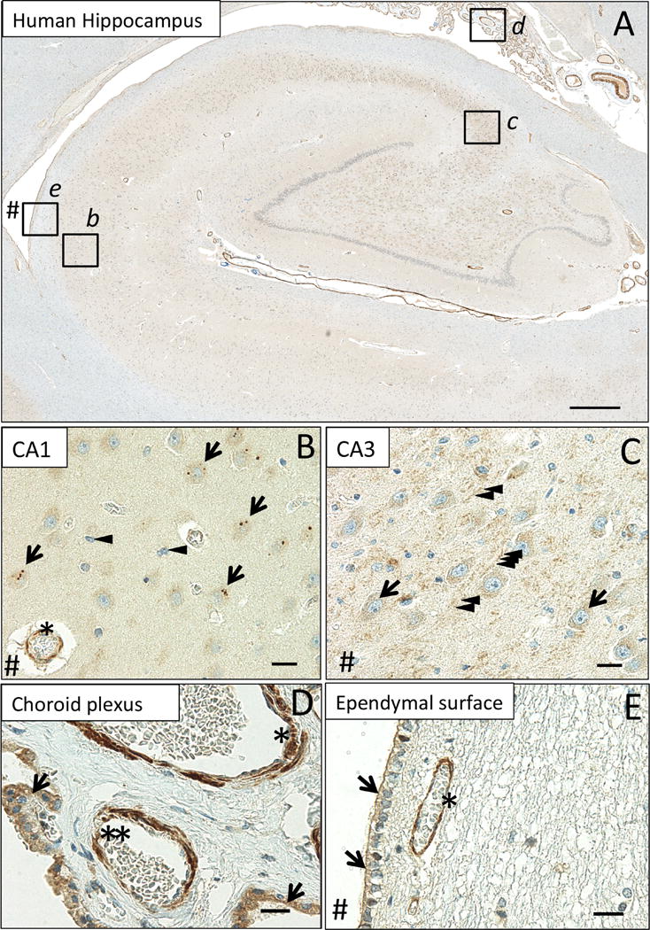
(A) MsrB3-immunoreactivity in CA1 (inset b), CA3 (inset c), choroid plexus (inset d) and the ependymal ventricular lining (inset e). In pyramidal neurons of healthy human (CDR0, BB0) CA1, MsrB3 was visualized as few somatic, sharply demarcated, 1-3 μm puncta (B, arrows); cortical glial cells do not give rise to MsrB3 signal (B, arrowheads). Asterisk indicates MsrB3-immunoreactive (-ir) cortical blood vessel wall. CA3 neuropil displays punctate MsrB3 signal (C, arrowheads) while CA3 pyramidal somata show diffuse signal (C, arrows). (D) The choroid plexus epithelium is MsrB3-ir (arrows). The thicker vascular wall of an artery (asterisk) and a thinner wall of a small vein (double asterisk) are MsrB3-ir. (E) Ependymal cells are MsrB3-ir at the apical surface (arrows) near an MsrB3-ir blood vessel wall (asterisk). # indicates (direction of) lateral ventricle. Scale bar A = 200 μm, B-E = 20 μm.