
Figure 7. MsrB3 signal in CA3 neuropil correlates positively with the percentage of CA1 pyramidal neurons exhibiting “healthy” MsrB3-immunoreactive phenotype (rare, ≤2, somatic puncta).
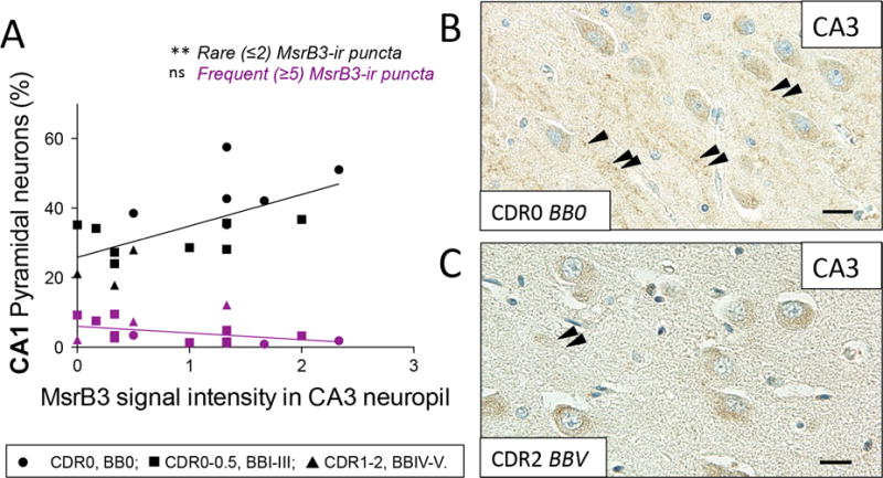
(A) The intensity of MsrB3 signal in CA3 neuropil positively correlated with the percentage of CA1 pyramidal neurons with rare (≤2) MsrB3-immunoreactive (-ir) puncta. (B) MsrB3 signal in CA3 neuropil in a healthy control (CDR0, BB0, arrowheads) compared to a hardly discernible neuropil signal in an AD subject (CDR2, BBV) (C, arrowheads). Scale bars = 20 μm. ns=nonsignificant, **p<0.01, by Pearson correlation.