
Figure 6.
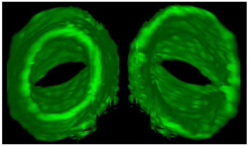
Two orientations of a 3D reconstruction of a lymphatic valve from a stack of confocal images. Left, looking downstream, into the narrowing bore of the valve between the leaflets, and at the outside of the vessel wall as it tapers outwards to the middle of the sinus. Right: looking upstream, directly at the free trailing edge of the leaflets and the twin blind fluid sacs formed between the inside wall of the sinus and the outer surface of the leaflets. From (Zawieja 2009).