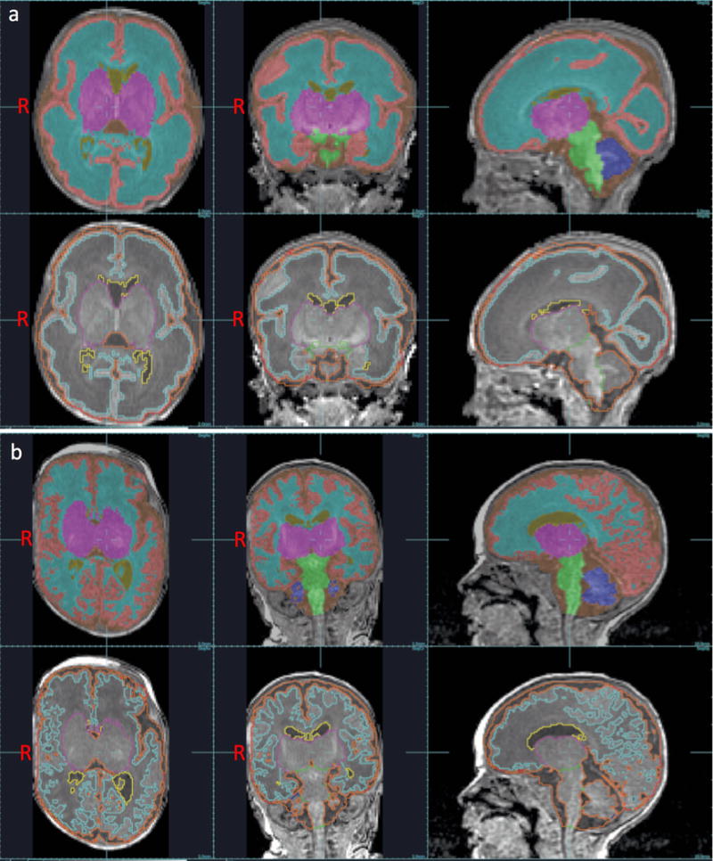
Figure 1. Brain tissue volumes at near-birth and near-term age.

Segmentations of cortical grey matter (peach), white matter (light blue), deep grey matter (fuchsia), cerebellum (dark blue), brainstem (green), and ventricular CSF (yellow) superimposed on 3D T1-weighted MRI images obtained near-birth (a), and near-term (b) age. Images are shown in axial, coronal, and sagittal planes.
