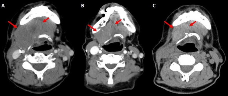
Figure 5. Local response to radiation.

(A) Computed tomography (CT) of the neck on October 28th, 2016. (B) Same performed on November 21st, 2016 after completion of three fractions of palliative irradiation, showing a reduction in the size of the main lesion as well as a reduction in size and numbers of adenopathy. (C) CT scan of the neck on March 28th, 2017. Unfortunately, IV-contrast was not used, so accurate assessment was difficult. However, the bulk of the disease had reduced in size.
