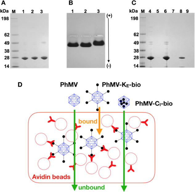
Figure 3.

Characterization of PhMV-biotin conjugates. (A) Biotinylated PhMV particles separated by denaturing SDS-PAGE visualized after staining with Coomassie. M = SeeBlue Plus2 molecular weight marker. (1) Native PhMV; (2) PhMV-CI-bio; (3) PhMV-KE-bio. (B) Biotinylated PhMV particles separated by agarose gel electrophoresis visualized after Coomassie staining. (C) Flow through and eluted biotinylated particles from avidin bead binding assay separated by SDS-PAGE and stained with Coomassie. (4) Native PhMV flow through; (5) PhMV-KE-bio flow through; (6) PhMV-CI-bio flow through; (7) bound native PhMV; (8) bound PhMV-KE-bio; (9) bound PhMV-CI-bio. (D) Avidin bead assay: PhMV samples are exposed to avidin-coated beads; only particles with biotin on the external surface bind to the beads.
