Figure 6.

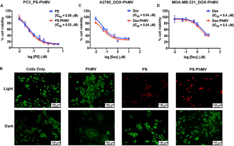
Evaluation of the cytotoxic efficacy of drug-loaded PhMV particles. (A) MTT cell viability assay of PC-3 cells using PS-PhMV. Cell viability was measured following 8 h incubation with varying concentrations of PS or PS-PhMV and 30 min illumination with white light (no cytotoxicity was observed when cells were incubated in the dark, not shown). (B) LIVE/DEAD assay of PC-3 cells showing representative images after photodynamic therapy of cells incubated with PS-PhMV or free PS and LIVE/DEAD cell staining. Calcein-AM staining of live cells is shown in green, and ethidium homodimer-1 staining of dead cells is shown in red. Scale bar = 100 μm. Illuminated cells incubated with PS-PhMV showed a slight increase in cytotoxic efficacy (IC50 = 0.03 μM) compared to free PS (IC50 = 0.05 μM). Dark controls show no cytotoxicity with PS-PhMV or PS. Scale bar = 100 μm. (C, D) Efficacy of DOX-PhMV (red line) versus DOX (blue line) using A2780 (human ovarian cancer) and MDA-MB-231 (human breast cancer) cells as determined by MTT assay. Cells were treated with DOX or DOX-PhMV corresponding to 0, 0.01, 0.05, 0.1, 0.5, 1, 5, and 10 μM for 24 h. IC50 values were determined using GraphPad Prism software.
