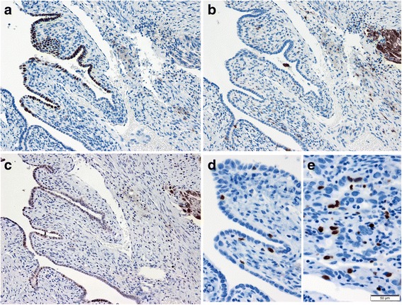
Fig. 2.

γ-H2AX expression in the distal fallopian tube. (a)-(c) Representative immunostained images of the p53 (a), p16 (b) and γ-H2AX (c) protein expressions in the high-grade serous carcinoma (right) and p53 signature (left). (d)(e) Representative Ki-67 immunostained images of p53 signature (d) and high-grade serous carcinoma (e)
