Fig. 3.

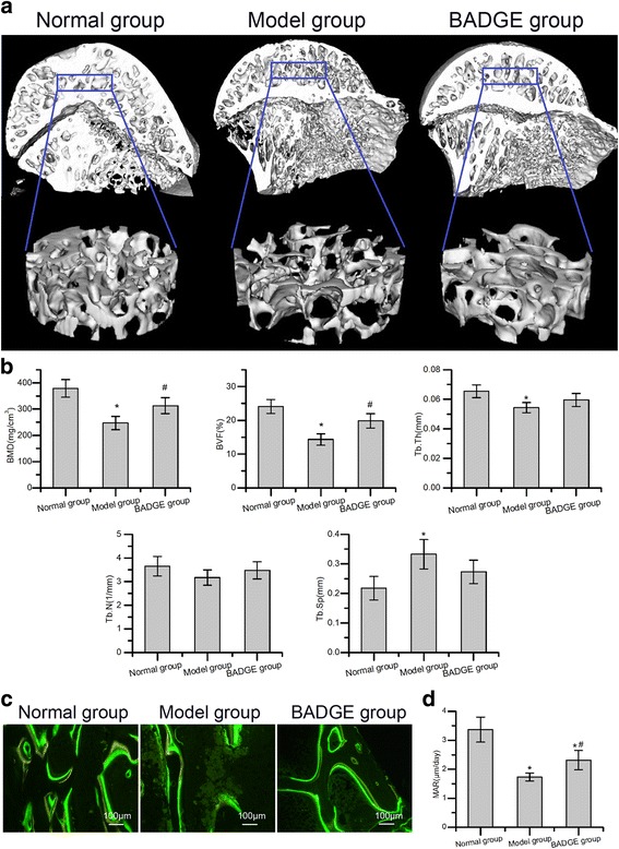
Treatment with PPARγ inhibitors ameliorated microstructural parameters and mineral apposition rate of trabecular bone. a Representative 3-D structure of femoral head of each group 6 weeks after the induction of osteonecrosis. b Quantitative analysis revealed significant reductions in BMD, BVF and Tb.Th and a significant increase in Tb.sp. in the model group compared with the normal group. Compared with the model group, the BADGE group exhibited significantly increased BMD and BVF (n = 5). c Representative tetracycline and calcein labelling images. d Quantitative analysis revealed a significantly increased mineral apposition rate in the BADGE group compared with the model group (n = 5). *P < 0.05 versus normal group; #P < 0.05 versus model group
