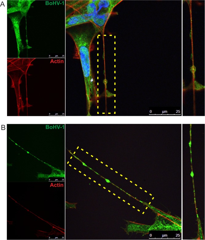
FIG 4.
Detection of BoHV-1 in tunneling nanotubes. KOP cells (A) and bovine fibroblasts (B) were infected with BoHV-1-WT (MOI = 1). At 12 h.p.i., the cells were fixed and stained. BoHV-1 proteins were detected using anti-BoHV-1 serum (green staining). Additionally, the cells were stained for F-actin (red staining) and nuclei (blue staining). Zoomed images of nanotubes (boxed regions) are shown at the right. Images were obtained using a Leica TCS Sp8 X confocal microscope.

