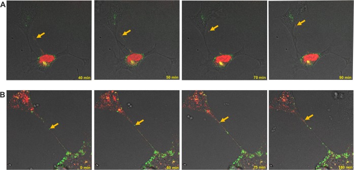
FIG 8.
Cell-to-cell transfer of dually fluorescent BoHV-1 mutant through tunneling nanotubes in the presence of neutralizing antibodies. KOP cells (A) and bovine fibroblasts (B) were infected with dual-color fluorescent mutant BoHV-1-gE-GFP-VP26-mCherry. Red, viral capsids; green, envelope glycoprotein E. Selected images from live microscopy recordings show fluorescently tagged structures moving inside the tunneling nanotubes (indicated by yellow arrows) from infected to uninfected cells (see also Movies S4 and S5). Images and time-lapse movies were obtained using a Leica TCS Sp8 X confocal microscope.

