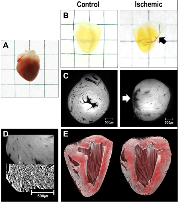
Figure 2.
Representative figure of tissue-cleared hearts and light-sheet images. (A) Sample of a mouse heart before tissue-clearing process (B) Cleared heart samples of control (left) and ischemic hearts (right). Suture used for the permanent ligation of left anterior descending artery can be seen (black arrow) (C) Light-sheet image of control (left) and ischemic (right) hearts. Infarcted area with thinning is observed (white arrow) (D) Image processing of raw light-sheet images (upper panel) using Sobel filters (lower panel) (E) 3D reconstruction of the entire heart from light-sheet images.

