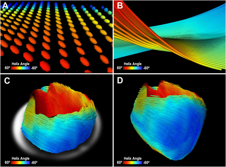
Figure 4.
Representative image of mouse heart DTI. (A) Zoomed-in view of 3D diffusion tensors calculated at each voxel in the short-axis plane. Orientation of the anisotropy of the diffusion tensor is conventionally assumed to be parallel to the underlying cardiomyocytes orientation. Diffusion tensors are color-coded based on the calculated helix angle. (B) Zoomed-in view of 3D cardiomyocytes tracking in the myocardium. (C) Reconstructed cardiomyocytes above the mid-left ventricle on a short-axis plane. (D) 3D tractography of the helix angle of a mouse heart visualizes the helical structure of the cardiomyocytes twisting around the left ventricle.

