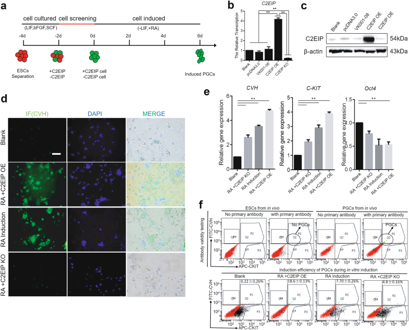
Fig. 3. C2EIP promotes PGC generation in vitro.
a Schematic diagram of RA induction model and C2EIP modification. Expression of C2EIP mRNA and protein in the different RA induction models was detected by qRT-PCR (b) and western blot (c), respectively. C2EIP OE and KO groups represent ESC transfected with C2EIP overexpression and knockout vectors, respectively. ESC mock-transfected with no plasmid were the blank control. d Fluorescence microscopy after 4–6 days showing PGC-like CVH-positive cells. ESC without RA induction was the negative control, ESC with RA induction and no transfection was the positive control, Scale bar:60μm. e qRT-PCR was used to quantify CVH, C-KIT, and OCT4 expression after C2EIP knockout or overexpression. f Antibody-specific detection of CVH and C-KIT by flow cytometry. ESC without RA induction was the negative control, and ESC with RA induction and no transfection was the positive controls

