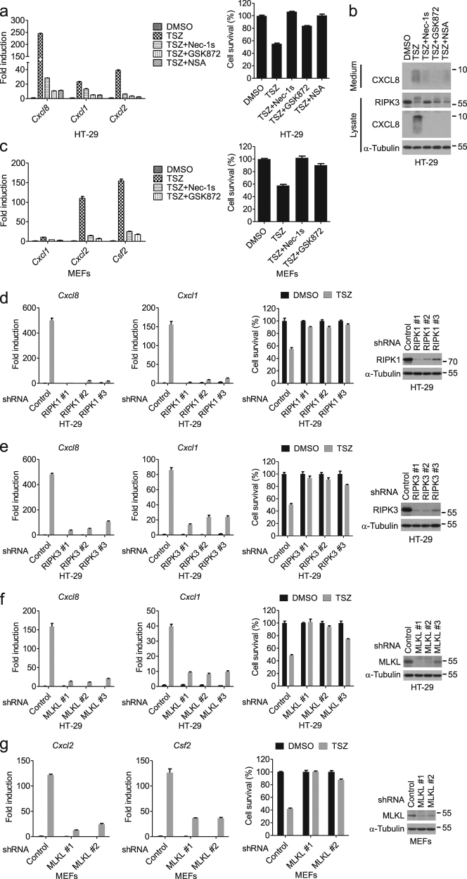
Fig. 3. The RIP1–RIP3–MLKL axis is required for the induction of cytokines associated with necroptosis.
a HT-29 cells were treated as indicated. The mRNA levels of Cxcl8 and Cxcl1 were measured by qPCR after 8 h of treatment (left). The cell viability was determined by CellTiter-Glo after 24 h of treatment (right). b HT-29 cells were treated as indicated for 8 h. Cell culture medium and cell lysates were collected separately, followed by western blotting analysis. c MEFs were treated as indicated. The mRNA levels of Cxcl1, Cxcl2, and Csf2 were measured by qPCR after 4 h of treatment (left). Cell viability was determined by measuring ATP levels after 13 h of treatment (right). d–f HT-29 cells stably expressing the indicated shRNA were treated with DMSO or TSZ. The mRNA levels of Cxcl8 and Cxcl1 were measured by qPCR after 8 h of treatment. The cell viability was determined by CellTiter-Glo after 24 h of treatment. The knockdown efficiency was determined by western blotting. g MEFs stably expressing the indicated shRNA were treated with DMSO or TSZ. The mRNA levels of Cxcl2 and Csf2 were measured by qPCR after 4 h of treatment. The cell viability was determined by CellTiter-Glo after 22 h of treatment. The knockdown efficiency was determined by western blotting. Data were represented as mean ± SEM of triplicates

