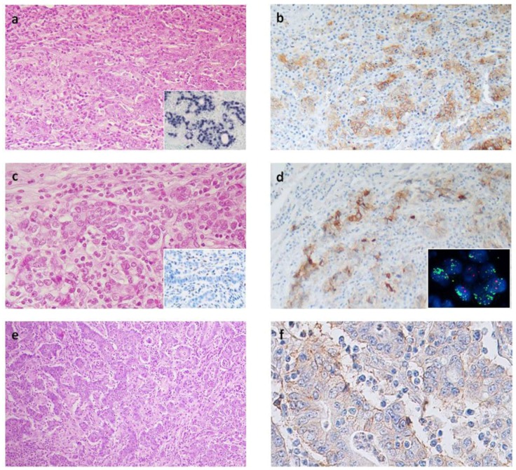
Figure 1.
(a) EBV+ gastric carcinoma showing lymphoid stroma and (b) abundant PD-L1+ tumor cells spread out along the tumor; (a inset), in situ hybridization for EBV RNA, EBER; (c) The MSI gastric cancer lacking MSH2 protein (c inset); with (d) intense PD-L1 expression prevalently along the infiltration front and (d inset) high levels of PD-L1 gene amplification; (e) One of the MSS/EBV− gastric carcinoma showing abundant lymphocytes infiltration and (f) weak PD-L1 immunoreactivity along membrane of tumor cells. Original magnification: a, b, d, e, a inset and c inset, 100×; c, 200×; f, 400×; d inset, 1000×.

