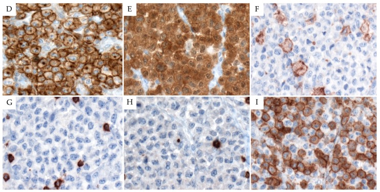
Figure 1.
Morphological and immunohistochemical findings in ALK+ ALCL. (A) Tumor cells infiltrating the sinuses displaying a cohesive pattern (H&E stain, 100×). (B) The large neoplastic cells are relatively monomorphic, showing abundant eosinophilic cytoplasm and pleomorphic nuclei and frequent apoptotic bodies (H&E stain, 400×). (C) A “hallmark cell”, displaying an eccentric horseshoe-shaped nuclei with two nucleoli and prominent Golgi area (H&E stain, 630×). (D) The tumor cells are strongly and uniformly positive for CD30 with a membranous and Golgi zone pattern. (E) Strong cytoplasmic nuclear and nucleolar ALK staining in the neoplastic cells is observed. (F) CD4 is positive in macrophages but negative in neoplastic cells. (G,H) CD3 and CD7 are positive in accompanying T cells but negative in the tumor cells. (I) CD5 is positive in the majority of the tumor cells ((D–I), immunohistochemistry, 400×). Abbreviations: H&E: hematoxylin and eosin.


