Figure 2.
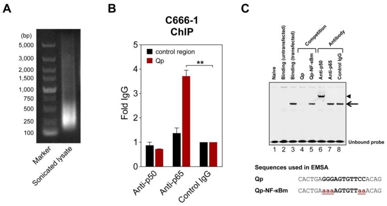
NF-κB can bind Qp. (A) Representative image of an agarose gel showing DNA present in a sonicated C666-1 cell lysate used for chromatin immunoprecipitation (ChIP) experiments. (B) ChIP of NF-κB p50 and p65 with Qp in C666-1 cells. Results of real-time PCR analysis ChIP assays with antibodies specific for p50 and p65 and a rabbit control IgG are shown. The results are expressed as fold enrichment, where the rabbit control IgG was set to 1. A genomic region ~5 kbp upstream of the IL-8 promoter was used as the negative control region. The means and SEM from three independent experiments are shown, and all samples were analyzed in triplicate. Statistical significance was calculated using the two-tailed Student’s t-test. ** p < 0.01. (C) An IRDye700-labeled DNA probe corresponding to the Qp region, spanning positions -83 to -63 of the B95-8 EBV sequence, was incubated with nuclear extracts from HEK 293T cells transfected with p50 and p65 and subjected to EMSA. Lane 2 shows the binding pattern of the nuclear extract from untransfected cells, while lane 3 shows the binding band associated with transfected cells, indicated by an arrow. Lanes 4 and 5 show the binding of the probe when an excess of a competitor oligonucleotide was added to the binding reaction. Super-shift experiments with antibodies were performed as indicated above the gel; the super-shifted complex is indicated by an arrowhead. Nucleotide sequences of the double-stranded probes and oligonucleotides used in the competition experiments are shown below the gel image. The NF-κB binding site in the Qp promoter is in bold uppercase letters, while the mutated nucleotides are in bold red lowercase letters and underlined. The gel displayed is representative of results obtained in two independent experiments.

