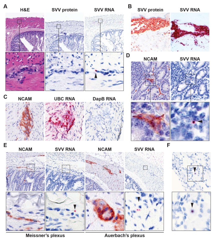
Figure 1.
Detection of viral RNA in enteric neurons of latently simian varicella virus (SVV)-infected rhesus macaques. (A) Consecutive intestine sections from latently SVV-infected rhesus macaques (RM) were stained with hematoxylin and eosin (H&E), immunohistochemically (IHC) stained for SVV nucleocapsid protein or stained for SVV ORF63 RNA by in situ hybridization (ISH). Note punctate red ISH staining (arrow head), indicative of low level ORF63 expression. Magnification: 100×; inset: 1000×; (B) Validation of SVV-specific IHC and ISH staining in varicella skin biopsies from SVV-infected African green monkeys [AGMs 273 (IHC) and 269 (ISH)]. Magnification: 200×; (C) Consecutive intestine sections from latently SVV-infected RM were assayed for NCAM expression by IHC, and RNA expression of ubiquitin C (human cellular UBC gene; positive control) and DapB (bacterial gene, negative control) by ISH. Representative images are shown for RM 9021. Magnification: 400×; (D,E) Consecutive RM intestine sections were stained for NCAM by IHC and SVV ORF63 RNA by ISH. Three SVV DNA qPCRP°S intestine paraffin blocks (each containing three distinct biopsies) from two RM (Table 2) were analyzed; (F) Representative images of a SVV ORF63 ISHP°S neuron in latently SVV-infected lumbar dorsal root ganglia of RM 2207; (D,F), magnification: 400×; (E) magnification: 200×. Inset shows enlargement of area indicated by the black box. Arrowheads indicate SVV ISHP°S neurons. Representative images of SVV ISHP°S enteric ganglia (on average 4.3 ISHP°S ganglia per section) are shown for RM 2207 (A,E); and SVV ISHP°S nerve fibers (on average 14.3 ISHP°S nerve fibers per section) are shown for RM 9021 (D).

