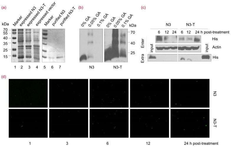
Figure 4.
NOP53-derived Protein N3-T Can Penetrate into Cells. (a) Lanes 1–7, His-fusion N3 and N3-T proteins were purified using a His-Ni column and the fusion proteins were analyzed by 12% SDS-PAGE. Lane 1, expressed vector control protein; lane 2, expressed His-fusion N3; lane 3, expressed His-fusion N3-T; lanes 4 and 5, protein molecular-weight markers in kDa as indicated; lane 6, purified His-fusion N3; lane 7, purified His-fusion N3-T; (b) Purified His-fusion N3 and N3-T proteins were treated with 0%, 0.05%, 0.1% GA for 15 min at 4 °C, followed by SDS-PAGE and immunoblotting with an antibody against His; (c) HeLa cells in T-25 flasks were treated with N3 (20 µg/mL) or N3-T (20 µg/mL) for the indicated times. The cells and culture supernatant were harvested for detection using an anti-His antibody; (d) HeLa cells in 24-well plate were treated with N3 (20 µg/mL) or N3-T (20 µg/mL) for the indicated times. The cells were fixed and reacted with antibodies against His. Nuclei were stained with DAPI. Images were captured using 10× objectives with an Olympus IX73 microscope.

