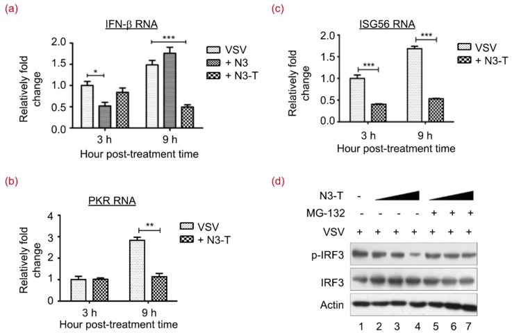
Figure 5.
N3-T Suppresses Type I IFN Antiviral Responses. (a–c) HEK293T cells were infected with VSV at 0.1 MOI for the indicated time in the presence of PBS, N3 (20 µg/mL), or N3-T (20 µg/mL). Total RNAs were isolated and expression levels of IFN-β (a); PKR (b); and ISG56 (c) mRNAs were quantified by real-time polymerase chain reaction (RT-PCR) and normalized to that of glyceraldehyde-3-phosphate dehydrogenase (GAPDH). The bar graphs show the means with standard deviations (SD) of triplicate repeats of one representative experiment. * p < 0.05, ** p < 0.01, *** p < 0.001; (d) HEK293T cells were infected with VSV at 0.1 MOI for 24 h in the presence of PBS or increasing doses of N3-T (0, 10, 20 µg/mL). The cells were treated with DMSO (lanes 2–4) or MG-132 (lanes 5–7) for 6 h before collection. Cell lysates were analyzed by immunoblotting with antibodies against p-IRF3 and IRF3. Actin served as the loading control.

