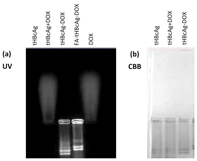
Figure 6.
Migration profile of the tHBcAg VLNPs conjugated with folic acid and doxorubicin in a native agarose gel. The same gel was (a) visualised under ultraviolet (UV) illumination and (b) stained with Coomassie brilliant blue (CBB). Samples labelled on top of the gel images are tHBcAg VLNPs (tHBcAg), tHBcAg VLNPs incubated with doxorubicin (DOX) without the conjugation steps (tHBcAg + DOX), tHBcAg VLNPs conjugated with DOX (tHBcAg-DOX), tHBcAg VLNPs conjugated with folic acid (FA) and DOX (FA-tHBcAg-DOX), and free DOX.

