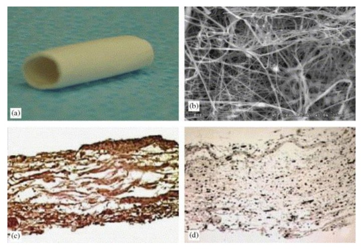
Figure 7.
(a) Scaffold before crosslinking; (b) scaffold before crosslinking at a magnification power of 1800×; (c) immunohistochemical analysis utilizing antibodies specific to collagen type I; (d) scaffold with 15% elastin shown a homogenous elastin network. Reproduced with permission from [119]. Elsevier, 2006.

