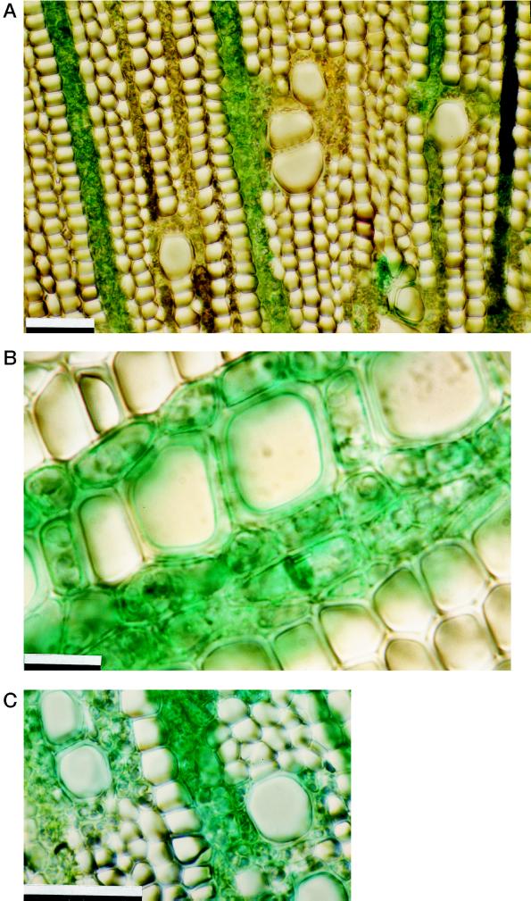
Figure 4.
Cross-sections of 1-year-old twigs in which Fast-Green dye was applied to the exposed cortex before pressurization. A, Green-stained rays showing that the dye was transported to the wood via the rays; B, detail of secondary wood (note green-stained multiseriate rays and parenchyma cells surrounding vessels); and C, detail of green-stained vasicentric parenchyma. Scale bars = 50 μm.

