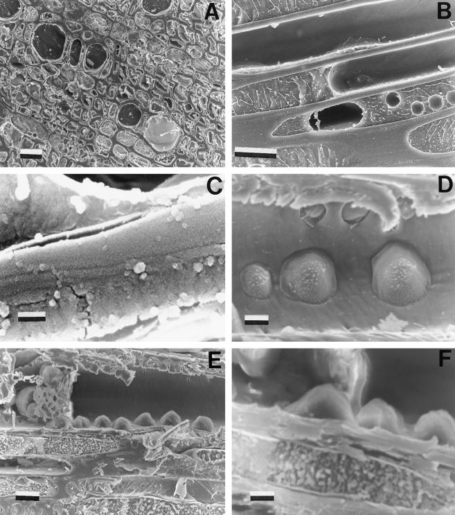
Figure 5.
A, Full (functional) and empty (dysfunctional) xylem conduits observed under a cryoscanning electron microscope. Twig samples were collected from prestressed plants. Scale bar = 100 μm. B, Cavitated vessel with a large bubble in it and a conduit with many small bubbles. Scale bar = 10 μm. C, Detail of a cavitated conduit showing the air/water interface and a sap layer persisting adherent to the conduit wall. Scale bar = 1 μm. D, Cavitated conduit at the beginning of refilling (note the numerous water droplets entering the conduit through the pits). Scale bar = 5 μm. E, Cavitated conduit at the beginning of refilling (same as D) showing the parenchyma cells close to the conduit. Scale bar = 10 μm. F, Detail of D at a higher magnification (note water droplets entering the conduit). Scale bar = 5 μm.

