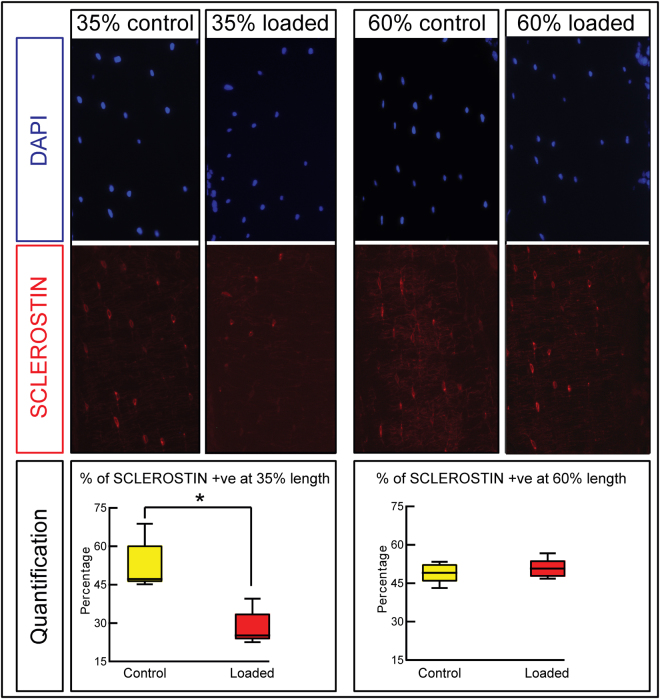
Figure 5.
Effect of excess strain load-priming on osteocytes’ SCLEROSTIN expression at 35 and 60% tibia length. 20 months old female mice in group 4 received two bouts of 11 N loading and sacrificed two days thereafter. Representative transverse DAPI and SCLEROSTIN-immunostained images with the percentage changes of SCLEROSTIN-positive osteocytes in non-loaded contralateral control and loaded right tibia demonstrating significant reduction in percentage of SCLEROSTIN-positive osteocytes at 35% of tibia length only. 8 sections from 4 mice per group were quantified. Box-plots represent means ± SEM. Group sizes were n = 4. *denotes p < 0.05.

